My Role as a SyncEquine Technician

SyncEquine-Cover

2 Years on ……

I have now been qualified and operating as a SyncEquine Thermal Imaging Technician for 2 years.  Cliche I know, but how time flies and there has not been a day I have regretted the decision to join the SyncThermology team.

Over the last 2 years I have met and worked with some lovely clients, professionals and indeed horses who I always find so endearing once they relax and take on their role of super model.

Sync’s team of vets offer excellent support and are always available to discuss and offer advice on each case.  As well as educating the team by giving presentations and live dissections at company CPD’s and team webinars.  I find each case fascinating and as my veterinary knowledge increases so does my passion for learning more.  If you are like-minded and would like to see how our screening service can benefit the equine veterinary industry.  Please read on!

In this blog I would like to share with you a handful of cases I have had the pleasure of being involved with.  Each being a reflection of some common veterinary issues I regularly come across along with some unexpected diagnosis’.  Soon after starting this blog I also realised are naturally linked to the company’s quarterly themes, in particular: Joint Disease, Pain or Behaviour, Poor Performance, Tendons & Ligaments and Detecting Early Pathology

Below are a summary of four cases, where I have pulled out the most relevant points from the SyncEquine Veterinary reports to share with you.

Case 1 August 2016 – Joint Disease/Poor Performance
My first client and scan I performed as soon as I had finished my training .  Myself and my client were both surprised to say the least at the findings.  We were both convinced there was something going on in this mare’s back, which we were not completely wrong,  But strongly reminded to not concentrate on looking at the symptom but to look closer for the cause.

History & Subjective Complaints:                                                       Logo SyncEquine Blue on White

  • 12 yr, Warmblood, Event horse, purchased to event at BE100 however has competed up to 1*
  • So far that season had unfortunately posed some issues for my client, in particular the XC phase due to refusals.
  • The rider had noticed that the saddle was not sitting straight but also that the mare’s hindquarters were not square.
  • The saddle was therefore checked and the mare has received  regular physio treatment from a registered Physiotherapist.  
  • Although there have been no other symptoms or lameness to cause any major concern my client wanted a fully body screening in order to check the whole body with the aim of finding an explanation to the poor performance.

SyncEquine Veterinary Report Highlighted:

Forelimbs:

  • Bilateral hyperthermia of the palmer metacarpal is observed on medial (inside) and lateral views at rest and post exercise L>R
  • Bilateral inflammation of the metacarpophalangeal joints pre and post exercise.
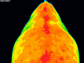
This slideshow requires JavaScript.

Body:

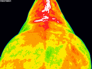
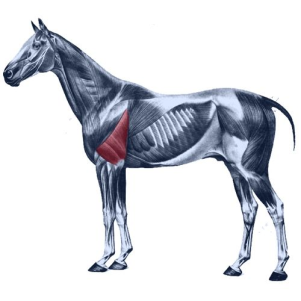
  • There is a distinct bilateral focus of inflammation on lateral views of the upper body in the caudal aspect of the lateral shoulder region, apparent post exercise.

This slideshow requires JavaScript.

Hindlimbs:

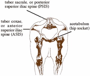
  • There is diffuse hyperthermic asymmetry of the left gluteal region on dorsal views of the hindquarters at rest
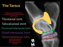
  • Focal intertarsal inflammation is observed on dorsal (back)) and medial views of the hocks at rest and post exercise
  • There is focal inflammation on the plantar aspect of the left tarsometatarsal joint
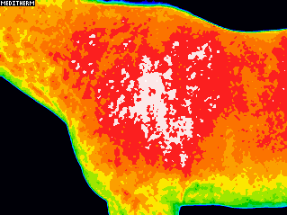
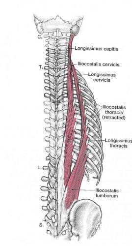
This slideshow requires JavaScript.

images

Spinal:

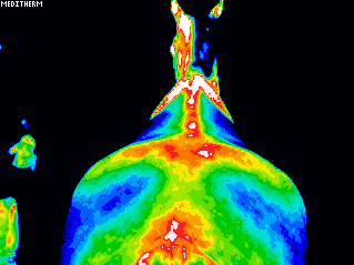
  • There is focal inflammation of the cranial thoracic vertebrae and left thoracic trapezius musculature – this is more apparent post exercise.

This slideshow requires JavaScript.

Discussion

  • Palmer metacarpal hyperthermia may not be significant in the absence of clinical signs of tendon or ligament pathology, these images may be used as a baseline for further monitoring
  • If positive to flexion tests, further clinical correlation of front fetlock and tarsal joint inflammation is advised. Such thermal patterns can represent degenerative joint disease and this should be investigated if there is clinical concern
  • Bilateral focal inflammation of the caudolateral shoulders is suspected to be secondary to saddle fitment and may explain saddle slippage
  • Left gluteal hyperthermia at rest is suspected to represent muscular tightness through this region
  • Cranial thoracic vertebral and left trapezius inflammation may also be linked to saddle figment. Spinal inflammatory patterns can, in some cases, be indicative of dorsal overriding of the spinous processes – however this would be unusual in the cranial thoracic region. This should however be investigated, if there is clinical concern.


Follow Up:
My client subsequently had her mare diagnosed further by her vet and the early onset of osteoarthritis was indeed suspected in the front fetlock joints.  Which was then treated with corticosteroid injections.  They together then went on to successfully finish the eventing season by going clear cross country.

During the winter the mare was also diagnosed with osteoarthritis in the both hocks and so my client decided to retire her from competing and concentrate on competing her other horses

Conclusion
There is no doubt in my mind that the cause of the poor performance was due to the mare experiencing pain in her front fetlocks.  I am also certain that the tightness in the mare’s shoulders and left hind quarters was secondary to osteoarthritis in the fetlocks and hocks.  The issue in the upper back may also have been linked but there was also the plausible explanation of the saddle fitting, which the owner had already addressed.

Case 2 June 2-17 – Pain or Behaviour
The reason the owner called for a consultation about her competition driving pony was to determine whether indeed the reason for the change in his behaviour when ridden was caused by pain.


History & Subjective Complaints:                                                 Logo SyncEquine Blue on White

  • Predominantly a competition driving pony, also ridden under saddle
  • Nov 16 – started bucking whilst ridden – physio identified a spasm in right hind quarter and left shoulder
  • April 17 – referral to vet, declared sound so to carry on working and have regular physio treatment
  • Barefoot but worked in hoof boots

Body:

  • Focal inflammation of the *CT is observed on right lateral views of the neck pre and post exercise (*Cervical Thoracic Junction – Neck to Upper Back)
  • No other significant thermal abnormalities observed.

This slideshow requires JavaScript.

Spinal:

  • Focal inflammation of the caudal thoracic spine is observed on dorsal views of the back pre and post exercise

Feet:

  • There is normal dorsal thermal symmetry of the forefeet and hind feet
  • On palmer views, there is medial and lateral solar hyperthermic asymmetry of the left and right forefoot respectively
  • Hyperthermic asymmetry of the right heel is observed on standing palmer views at rest, this is normal post exercise

Discussion:

  • There is thermal evidence of mild foot imbalance in the forefeet – this improves after exercise. Thermal evidence suggests that there is a tendency to lean to the right and increase weight bearing on the right forefoot
  • An assessment of the neck is advised – focal inflammation at the level of the CT junction may indicate muscular or spinal dysfunction at this level
  • Inflammation of the caudal thoracic spinal region warrants further clinical correlation as this thermal pattern may represent muscular tightness through this region, spinal ligamentous sprain, or in more serious cases – dorsal overriding of the spinous processes (kissing spine)


Follow Up, 1 Week Post Scan
Feedback from Client following Owner’s Vet Assessment: 

OLYMPUS DIGITAL CAMERA

  • Full workup performed and observed as sound on both hard and soft going
  • However in canter in the arena, very tight, rigid back with no movement
  • Evidence to justify x-raying the back
  • X-ray showed there to be no areas of concern in the lumbar region with regular even spaces between each of the spinous processes
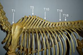
  • The thoracic spine looked good with the exception of an impingement between T10 & T11.
  • Vet did also note that there was a lot of tension in right side of Alfie’s neck (T1 area)

Follow Up 2 Days Later:

  • Nerve blocked offending vertebrae (T10 & 11)
  • The change in way of going was significant, much more free through the shoulder, worked with a much longer stride, and easily maintained left canter
  • Still looked tight over the sacroiliac, but with the amount of tension and spasm he had there it’s not surprising

Follow Up 2 Weeks Later:

  • Surgery performed (a wedge oseteotomy) being the best option
  • Injecting with steroid may have given short term relief but wouldn’t resolve the problem long term
  • Also had mild laminitis when given prednisolone to treat a respiratory infection a couple of years ago, so it would be too risky to medicate with steroids again.
  • 3 -4 month rehab program, but hopefully this should make him a lot more comfortable and resolve the neck issue too

Four Months Later: 

Significant improvement and successfully returning back to competing!

Conclusion
As owners we obviously know our equine partners very well as we work with them and see them on a regular basis.  So if your horse’s behaviour changes of course it makes perfect sense to not ignore it but to investigate further.  At the very least to rule out whether your horse is in pain.

In this case yes the pony was clearly in pain which due to the location of the impingement in the thoracic spine, was not surprisingly more noticeable when ridden and when the pony showed his discomfort by bucking.  However as vets it is very difficult to make a diagnosis especially when there is no obvious lameness or discomfort.  But as owners we also have to follow our gut when we know that our horse just isn’t comfortable.   This is when our screening service proves an excellent tool to isolate areas of pain.  Effective even more when used in conjunction with the vet’s diagnosis.   Which in this case was literally a pain in the neck, no doubt connected to the upper back (T10 & T11) which in turn would explain the tightness over the sacroiliac.

Case 3 October 2017 – Tendon & Ligament

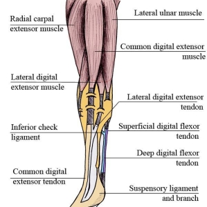
Injuries are common in the key tendons which run like cables down the back of the lower leg (deep and superficial digital flexor tendons). These tendons extend from muscles high in the leg to the foot.  When the muscles contract, they flex the leg. They also help support the leg and come under huge stress when a horse gallops, jumps or does any sort of athletic maneuver.

In the following case, the client’s vet suspected damage to the left fore suspensory ligament.  However as the nerve block was inconclusive he endorsed using our screening service to see if we could pinpoint the area of concern.

History & Subjective Complaints:                                                     Logo SyncEquine Blue on White

  • 10 yr old, TB, Ex Racehorse
  • slipped in the field about 8-10 weeks ago, evidence of which could be seen by skid marks and a broken fence
  • After which he did not feel or look quite right
  • The vet attempted nerve blocking but could not block sound and no difference was seen
  • However suspected damage to the origin of the suspensory ligament (where it attaches to the back of the knee)
  • Vet recommended six weeks of box rest and three sessions of shock wave over two weeks
  • During the walking exercise of the rehab programme, his owner noticed that he didn’t look sound
  • Vet re blocked behind the knee which resulted in improving soundness but still not 100%
  • Recommended now to have further diagnostics, X-Ray, Ultra Sound and MRI

Forelimbs:

  • Significant focal intercarpal inflammation is observed on dorsal views of the left carpus pre and post exercise. On lateral views there is diffuse hyperthermic asymmetry of the left carpus
  • A small focus of inflammation is observed on lateral views of the *proximal left metacarpus
  • On palmer carpal views, there is focal inflammation of the medial aspect of the radiocarpal joint

* Proximal – closest to

Discussion:

  • There is no thermal evidence of pathology at the proximal attachment of the suspensory ligament
  • The primary thermal finding is inflammation of the left dorsal intercarpal region with diffuse generalised hyperthermic asymmetry of the left carpus
  • There is also focal inflammation of the left metacarpus, *distal to the carpometacarpal joint – Further imaging to determine the cause of inflammation is recommended

* Distal – farthest away

Follow Up:: October 2017
Examination – diagnostic Testing, Radiographs L Carpus & Ultrasonography:

Information for Owner
Our examinations have identified a multi-site lameness, partially referable to the carpus (knee) and the carpal sheath, where a lesion in the superficial digital flexor tendon has been identified.

Information for Referring Vet
The diagnostic imaging findings are in agreement with the results from diagnostic analgesia. (nerve block)
6e0a167da31ce70c23be0637f2c2b083544e956f

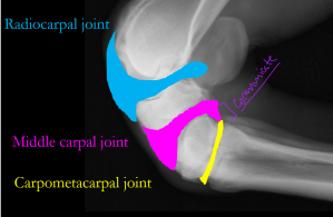
Whereas we think that part of the lameness originates from the (distended) LF middle carpal joint 

74028542e8add9832fe53a59c28b98a6
The tear in the LF SDFT is likely to be contributing to the lameness more substantially.

Ultrasonography:

Archie Crowther
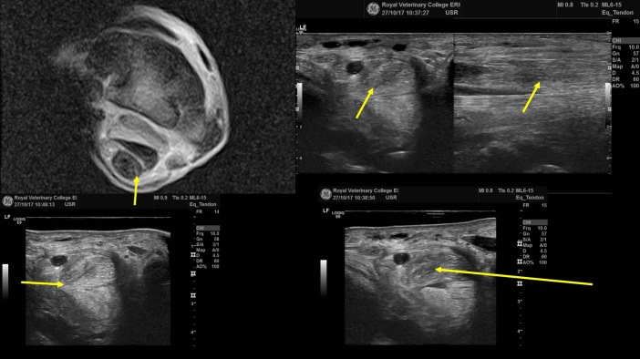
  • The bone surface at the origin of the left fore suspensory ligament appeared irregular (enthesopathy), but the size and fibre pattern of the ligament was within normal limits. The lesion within the left fore SDFT at the level of the left fore superficial digital flexor tendon was identified as a distinct area of hypoechogenicity and altered fibre pattern distal to the musculotendinous junction and was associated with a partial transverse split of the tendon at this level – Diagnosis: Superficial digital flexor tendinitis

Conclusion:
This case resulted in complete correlation with the client’s vet and the subsequent referring equine clinic.

  • The SyncEquine report highlighted inflammation of the left dorsal intercarpal region with diffuse generalised hyperthermic asymmetry of the left carpus.
  • The equine clinic agreed that part of the lameness originated from the LF middle carpal joint.
  • The SyncEquine report also identified a focal inflammation of the left metacarpus, *distal to the carpometacarpal joint.
  • The ultra sound identified a lesion within the left fore SDFT at the level of the left fore superficial digital flexor tendon.  (A distinct area of hypoechogenicity and altered fibre pattern distal to the musculotendinous junction was associated with a partial transverse split of the tendon at this level).

To find out how our screening service can detect ligament and tendon damage before reaching an acute level read more in next case

Case 4 Feb 16 & March 18 – Early Pathology
I was asked by Head Office if I could perform a follow up full body scan on this mare.  The client’s veterinary practice were so impressed with our initial findings in February 2016 that they were keen to share this case with other veterinary practices.  This was certainly another interesting case which further evidences the effectiveness of thermal imaging as an early diagnostic tool.

History & Subjective Complaints:                                                   Logo SyncEquine Blue on White

  • Owner reported tendencies to ‘nap’ when turning on a tight circle to the left
  • When jumping she also favoured the LF on landing.
  • There were also some additional areas of concerns unrelated to the LF so a full body scan was completed.

Numerous areas of interest were reported on but the most significant finding was identified:

Forelimbs:

  • inflammation of the RF foot. Inflammation significantly increased post exercise on dorsal and palmer views

Discussion:

Assessment of the feet to rule out deep foot pathology was advised by our interpreting vet, alongside other observations.

Follow Up:
At the time of the first scan in February 2016,  was sound so the findings in the RF were not considered significant by her vet.  However in May 2016 the owner noted she was not comfortable weight bearing on the RF for extended periods of time, for example when putting studs in. In June 2016 Nimmy became lame in front, seemingly both front feet. Corns/bruising was suspected but it didn’t improve with treatment. Nimmy’s owner arranged for her front feet x-rayed in July 2016, these were clear so a period of rest followed. By mid August 2016 she appeared sound so gentle exercise commenced. After a successful start she became lame again in November 2016 following her first dressage competition. The treating vet completed nerve blocks which were inconclusive so in December 2016 an MRI of the forefeet was completed. MRI identified a 10 cm lesion of the RF deep digital flexor tendon (DDFT), the LF was normal.

Images show T1 HR transverse scan of the right fore foot showing a core lesion of the medial lobe of the deep digital flexor tendon.

March 2018 Report

History & Subjective complaints:

  • The reason for the scan is as a follow up to a SyncEquine Full Body scan which was performed in February 2016, where subsequent veterinary report where deep foot pathology was identified in the Right Fore
  • Further investigation via MRI in December 2016 detected a 10 cm lesion in the RF DDFT
  • The owner requested a second SyncEquine scan to assess the current situation and then regular thermography scans to monitor progress and check coping with the rehabilitation plan

Forelimbs:

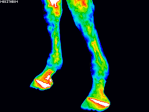
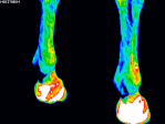
  • Focal hyperthermic asymmetry of the palmer right metacarpophalangeal and pastern region is observed on lateral and palmer views at rest and post exercise.

Pre Exercise:

Post Exercise

Discussion:

  • Significant hyperthermic asymmetry of the palmer right fore fetlock and pastern is likely to represent on-going tendonitis
  • Thermal findings in the right forefoot are suggestive of on-going deep foot pathology (navicular?) but hyperthermia could be secondary to tendon inflammation
  • Left forefoot solar hyperthermic asymmetry post exercise may be the result of lateral weight transfer from the right forelimb

Follow Up:
FURTHER INVESTIGATION: In January 2017 a RF Navicular Bursoscopy was performed  revealing a synovial mass in the medial navicular bursa, associated with a long split of the medical lobe of the tendon and an erosion of the lateral lobe of the deep digital flexor tendon. The synovial mass was removed and the torn fibres debrided from the DDFT. Bone marrow was also collected for culture of stem cells which would be injected in the Navicular Bursa 1 month after surgery. Following a period of box rest, then in hand walking and restricted turnout, allowed unrestricted turnout in April 2017. Further stem cells and steroids were injected over July/August 2017, however a routine appointment found the horse still 1-2/10 lame in November 2017 so an MRI was booked for December 2017 which showed guarded improvement.

Conclusion:
Pathology was detected with physiological imaging during the early stages of investigation when the patient was presenting subtle changes. Isolating abnormality for further investigation was a success.


Summary
I hope you have found each of these cases as interesting as I have.  For me the most satisfying aspect of my job is when my cases correlate with veterinary findings.  What is even more rewarding is when my cases are added to the company’s case study portfolio.  Which is constantly growing with more cases that evidence further the technology we use and the way we use it, certainly is proving it’s worth as a tool for the diagnostic tool kit.  Along with being used for pain evaluation and treatment monitoring, of course benefits the equine veterinary industry by often resulting in being more cost effective to the owner, less stress for both the horse and owner and a much speedier diagnosis for all involved.

I will be sharing with you more cases in future blogs but in the meantime please make sure you like and follow our Facebook page (https://www.facebook.com/syncequine/) where you can see further cases which myself and other technicians are involved with.

If you are interested in finding out more about what our service offers or would like a free consultation please do not hesitate to contact us through our website: http://www.syncthermology.com


Alone we can do a little. Together we can do so much.

I first met Rob Jackson, the Horseback Vet in 2016 when he came to assess and treat one of my livery yard owner’s horses following me performing Digital Infrared Thermal Imaging (DITI) as a newly qualified SyncEquine Technician.

b5d67709f2d9c6bfdea5e20c25a25ad8

Merehead, an 8 yr old French TB currently being re trained as an eventer following a racing career had been displaying signs of intermittent bilateral hind limb lameness. On occasions it looked more like stiffness than lameness as the work was increased, becoming more noticeable and appearing uncomfortable.  At this time Merehead was also looking noticeably poor and generally not happy but with no obvious symptoms to pin point what the problem could be.  So SyncEquine was selected as the initial investigation of choice before continuing with further clinical investigation.

Merehead was one of the first horses I scanned following my training and what a fascinating case to start with!

The SyncEquine veterinary report that followed Merehead’s thermograms highlighted:

‘Thermal findings in the hindquarters may correlate with a history of intermittent bilateral HL lameness. Sciatic groove hypothermia and SI hypothermia are indicators of pelvic dysfunction which can lead to HL lameness – a pelvic assessment is advised’


Pelvis

Rob was recommended to the owner through her saddler and luckily she managed to get him out quickly (for those of you who know him, he certainly is in huge demand)

As soon as I knew Rob would be coming out to assess Merehead, I sent our veterinary report to him with of course an introduction from myself and an explanation of how SyncEquine operates.  Part of our service always involves informing the client’s vets and other professionals that we plan to scan their client’s horse.  We then forward them a copy of the subsequent veterinary report.

Luckily for me Merehead’s owner could not be there when Rob arrived so asked if I could be there instead. I am so glad that I was able to, as in the all the years I have spent around horses and seen how different professionals work, I can honestly say I have never seen anything quite like this.

Rob fully embraced our veterinary report and was super complimentary about thermography as a diagnostic tool.  Rob himself has in the past has used thermography as part of his assessments and fully endorses it’s usefulness.

At first the process started off fairly standard with Rob wanting to see Merehead being trotted up.  Rob noted that there was asymmetry in Merehead’s pelvis and substantial tightness along the lumber region and hindquarters.  All of which Rob fully concurred with our thermograms and veterinary report.

Rob was like a breath of fresh air, breezing in and out, in between performing the most extreme osteopathic treatment I have ever seen let alone heard of!  I could literally hear things going pop and crack as I watched Rob move from one leg to another which he pulled up, down and out at angles I didn’t think were possible.  All of which he did calmly and quietly with a wonderful rapport.

Following the treatment Rob watched Merehead being trotted up again, and it was straight way obvious that the movement was a lot looser and freer across the pelvis.

Two years on and Merehead is now enjoying his life as an eventer, his owner has brought him on slowly and carefully, he looks amazing and is confidently competing at BE90 with a view to be moving on up to 100 at the end of this season.

34560311_2159563257605022_8319052726923165696_n  Merehead and Emma Brinkworth, BCA BE90, June 18

It was about a year later when I again worked alongside Rob, he was visiting another one of my client’s yards where he was going to assess and treat three horses.  Of course, I jumped at the opportunity and Rob was keen to see the thermograms which would help confirm his assessments.

Client 1- Rob’s Assessment:
Stiffness through shoulders due to being on the forehand and pulling himself along through the shoulders and lower back.  Which has subsequently caused the muscles in the quarters to not work correctly.

Mojo_LI
SyncEquine Thermogram showing asymmetry across the thoracic spine and sacroiliac area


Client 2- Rob’s Assessment:

Tight across around the withers and base of neck and blocked through both shoulders

The following SyncEquine Thermograms showing the above:

Pre Exercise                                      Post Exercise


Client 3 –
Rob’s Assessment:
Moderate-marked bilateral triceps spasms, latissimus dorsi hypersensitivity, pre-scapular hypertension and tight caudal withers.

Leo
SyncEquine Thermogram showing Latissimus Dorsi Hypersensitivity
and Tight Withers

The further two cases below are the most recent horses which both myself and Rob have worked with:

Clover – Rob’s Assessment:
Moderate bilateral triceps spasms and latissimus dorsi hypersensitivity. Tight withers and mild pre-scapular hypertension.

Clover
SyncEquine Thermogram showing  Latissimus Dorsi Hypersensitivity and Tight Withers


Ettie – Rob’s Assessment:
Moderate bilateral triceps spasms, and latissimus dorsi hypersensitivity. Slightly tight throughout longissimus thoracis. Mild bilateral longissimus lumborum.

Triceps     Ettie

Above – Diagram of Triceps Muscle and SyncEquine Thermogram

Ettie Back Diagram   Ettie 2

Diagram and SyncEquine Thermogram showing longissimus thoracis and longissimus lumborum


What was lovely about all of these cases was that the owners not only soon recognised there was a problem but were quick to investigate and take the advice from the SyncEquine veterinary reports.

It was fantastic to work together with Rob and see that the thermograms I performed correlated exactly with what Rob felt and saw.  It was also great to see that working holistically as part of a team with other practitioners really can give the best outcome for the horse.

After all this is one of the reasons I am passionate about my job as SyncEquine’s approach to the equine industry is unique. The aim being to ensure our service is used in every day veterinary medicine just like other diagnostic tests. In most cases thermography should not be used as a standalone diagnostic tool, it needs to be integrated into veterinary practices to be used effectively as a diagnostic test and prognostic indicator.

But for me personally I simply just want to see happy,  healthy horses and unworried owners!

For more information on SyncEquine please visit:
http://www.syncequine.com

You can also follow us on Facebook for regular updates on cases, research, training etc:
https://www.facebook.com/search/str/syncequine/keywords_search

More information about Rob Jackson, the Horseback Vet can be found at:
http://www.horsebackvet.com/about-us.html


SyncThermology: Soft Tissue Injuries and Baseline Imaging

I have had many falls that I have bounced back from but for the first time ever after suddenly and heartbreakingly losing my event horse of a lifetime ‘Rou’ back in RMW_2064{Class 1 British Novice.jpg
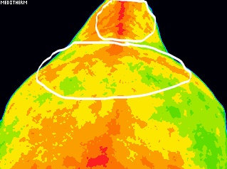
September.   I really was on the verge of giving up riding all together.  However, my wonderful trainer who is also a great matchmaker introduced me to a little ex racehorse who was mine to have on loan to see how we get on.   Well it has turned out that we get on very well.  So much so that I am truly grateful to this little mare for returning my mojo.

I have loaned Robyn since November and as soon as I started riding and getting to know her I realised this mare has something special about her.  She has a great training attitude that I find TB’s tend to have, absolutely loves her jumping and is such a pleasure to handle.  Being Irish and a mare what more can she have going for her?

blogI always need a goal to keep me focused, whether achievable or not and a training plan to follow.  So after swiftly changing my mind about giving up, I have kept busy schooling and training with Robyn, including the occasional show jump, dressage and arena eventing competition.  So far nothing has fazed her and as she is also proving to be hugely talented with a massive heart.  I decided to hell with it and register her BE this season.

A big part of my training plan for Robyn has to include monitoring her health and fitness which after all is the best defence against injuries.  I want my horses to always happily jig home sound, fit and uninjured every time they are ridden and especially after a competition.

I had noticed a couple of weeks ago, Robyn seemed sore around the right side of her withers so I asked my saddler to check her saddle.   In my saddler’s words “your horse has developed a fab back end” that had popped her saddle out of balance which my saddler soon rectified.  Robyn has also had a physio session where the physio found some tightness but nothing to cause too much concern.  Maintenance physio sessions will continue regularly throughout the season for both Robyn and myself.

SyncThermology’s focus this quarter on ‘Soft Tissue Injuries and Baseline Imaging
fits in perfectly with my competition plan for this season.  Baseline imaging is another excellent tool to use as a way of monitoring your horse’s health.  Therefore, perfect to use at the beginning of the season.  The imaging scans are kept on file with SyncThermology’s team of vets as a way of benchmarking a healthy horse and then used as a comparative for future scans.  For Robyn this is perfect as I can keep a check on the areas that I know she has slight soft tissue damage as well as using the technology to quickly identify any future lameness, conditions or concerns.

I have therefore taken a baseline Imaging Scan of Robyn, which has given me some peace of mind that I have invested wisely into her health for this year’s event season.
2017 – Bring it on!

blog 2

Equine Joint Disease

iStock_48086140_MEDIUM-1.jpg
Veterinary DITI, Assisting Animal Diagnosis, Treatment and Rehabilitation

In this quarter SyncEquine and SyncCanine are focussing on joint related issues.  This theme could not be more relevant to my most recent case and so will be the subject of this blog as well as explaining more about my role as a SyncEquine Imaging Technician.

The owner asked me to perform a full body scan on her 12 yr, event horse who she bought last winter to compete the following spring.   The mare has previously competed to BE 1* but as this is a new partnership they have so far been competing at BE100 this season.

Part of the service that SyncEquine Imaging Technicians provide to our clients ensures that the screening process is always performed in a professional and methodical manner.  The full body scan including the head comprises of recording uniform lateral, caudal and cranial sections of the body, (including 360-degree angles). The horse is scanned at rest (for at least 1 hr) and then again after 10 mins of moderate exercise (ideally lunging). This ensures that every report which is produced by our team of vets is as comprehensive and accurate as possible.

We advise that the first appointment be a full body screening especially if it is unclear if the issue is due to pain, behaviour or the horse has a nonspecific and difficult to diagnose lameness.  Once a holistic diagnosis has been made we recommend regular region of interest scans to monitor the progress.

So far this year has unfortunately posed some issues for my client, in particular the XC phase due to refusals.  Recently the rider has noticed that the saddle is not sitting straight but also that the mare’s hindquarters are not square.  The saddle has been checked and the mare receives regular physio treatment from a registered Physiotherapist.

Although there have been no other symptoms or lameness to cause any major concern my client wanted a fully body screening to explore the hindquarters further as well as having a full body health check.

The following pictures are of the cranial and lateral scans taken of the front legs section (below knees) of my client’s mare which are most significant to this blog.

Pre-Stress

Post-Stress


Summary of SyncThermology’s Vets Report:

  • Inflammation within the shoulders which is suspected to be due to saddle fitting (may explain saddle slippage)
  • hyperthermia within the left quarters (gluteal) which is suspected to represent muscular tightness through this region.

But what I found most interesting was the vet recommending my client’s vet to flexion test her mare and if positive:

  • further clinical correlation of front fetlock and tarsal joint inflammation is advised. Such thermal patterns can represent degenerative joint disease and this should be investigated if there is clinical concern.

My client subsequently had her mare checked by her vet and the early onset of osteoarthritis was found in the front fetlock joints.  The vet then treated the mare with corticosteroid injections.
Impact on Joints

Being the horsey geek I am I do find the anatomy of the horse fascinating and am not surprised to see the issues that commonly effect our performance horses, especially after studying the photo sequence below by Eadweard Muybridge.  The report from SyncThermology’s team of vets reminded me of an article I have read by Dr Robert Bell and Professor Leo Jeffcott, who are based at the Equine Performance and Imaging Centre, University of Sydney Veterinary Teaching Hospital Camden.

The following excerpt is taken from a report on ‘Fetlock Lameness – It’s importance and how MRI can assist in making the difficult diagnosis’ which was first published in 2011.

Lameness involving the fetlock joint is an all too common problem in performance horses and racehorses. Injuries to this region may involve the joint itself or the surrounding soft tissues, and are often determined by the use of the horse. The fetlock is a complicated high motion joint that is always subjected to huge forces and stresses during locomotion. The classic movie pictures of the American photographer Eadweard Muybridge taken over 120 years ago (pictured below) clearly show the degree of extension of the fetlock during galloping in the horse.

V8000048-High-speed_sequence_of_a_galloping_horse_and_rider-SPL.jpg
  Picture courtesy of Science Photo Library

The good news is that many fetlock problems are fairly simple to diagnose. Lameness can be localised to the fetlock by careful clinical examination (i.e. palpation), evaluation of gait (especially under saddle), response to flexion tests and nerve blocks or desensitisation of the joint itself.
http://www.horsemagazine.com/thm/2016/04/fetlock-lameness-its-importance

Monitoring

I am looking forward to monitoring my client’s horse progress and what will be most interesting to see is whether the mare’s performance during cross country improves.  If so there is no doubt in my mind that the problems were caused by the mare experiencing pain in her front fetlocks.  A question I also hope to see answered is whether the tightness in the mare’s left hind quarters is secondary to the osteoarthritis?  If there is an improvement I am sure the tightness in the left gluteal muscle was due to the mare carrying herself in a way so as not put too much weight on her front legs.

To Conclude:

Since the report I referred to above by Dr Robert Bell and Professor Leo Jeffcott was written in 2011 the case I have shared with you has also demonstrated the importance of Veterinary DITI.  Especially in this case the joint condition was certainly detected before it caused any lameness.  My experience of SyncEquine’s scanning process and seeing the SyncThermology vet reports for the horses I have scanned has proved that the technology we use and the way we use it is most definitely an effective and invaluable diagnostic tool.  The camera can certainly detect where the cause of the problem lies, identify early what the issue is and then monitor the progress of the horse.

In my future blogs I will share with you the progress of this particular mare as well as continue to share with you my experiences of being an Equine Technician for SyncThermology.

In the meantime please feel free to contact me with any questions or if you are interested in having a free consultation.

Louise Crow
SyncEquine Imaging Technician
lou@synequine.com
www.syncthermology.com


SyncEquine Imaging Technician

I have really enjoyed my training with SyncThermology which is now all complete and I’m all set and raring to go with my new business venture.

I am therefore proud to announce that I am now a fully trained SyncEquine Imaging Technician.

The reason I chose to pursue a career within this arena is due to redundancy and the ‘muck heap moment’ I had mainly because of the issues I have experienced with my event horse Rou, as explained in my first blog.

Because my role within children’s services involved recognising health and behavioural needs by working holistically to prevent, intervene and support vulnerable families.  I believe these themes can be echoed within the equine industry where prevention, monitoring, effective early diagnosis and rehabilitation are also imperative both for the client and more importantly for the horse.

SyncThermology specialises in DITI (Digital Infrared Thermographic Imaging) which research has proved an effective tool to early identify musculoskeletal and neurological injuries, in particular non-specific and difficult to diagnose lameness.

After much research I discovered SyncThermology not only specialise in clinical thermal imaging but offer a unique way of working within this industry. My experience of working within Children’s Services fit perfectly into SyncThermology ethos and allows my skills to be used in the most logical and effective way.  Working closely with the client alongside their vets and other practitioners as well as SyncThermology’s own team of vets provides the opportunity to work holistically to maximise the benefits of the technology for the patient.

But that is not all, the service I now provide involves performing dynamic testing which also makes our company unique from others and enhances its effectiveness.  The cameras we use for screening are of a clinical standard and are also used in the health sector in particular used to detect and monitor breast disease.

Part of my training entailed undertaking my own case studies and after discussing the concept with a fellow livery, she was really keen for me to use her daughter’s pony as one of my cases.

Training Case Study:
Pony was purchased as a Christmas present after her daughter completely and utterly fell in love with him.  Not long after ownership, pony became more and more nappy and generally not looking happy within himself when ridden.  The vet’s examination (determined by process of elimination) diagnosed the issue was within the sacroiliac joint and so the joint was injected with steroids and Harvey has shown great improvement after a period of rehabilitation. 

The data I collected was sent to the team of vets who analysis and provide veterinary reports as part of the service.  The report was comprehensive and certainly picked up the reason for the issues which is sacroiliac but also showed potential issues in the right hind foot and both hocks.  Harvey will continued to be screened to monitor his progress and hopefully ascertain that these are secondary implications. .

Already my own experience of DITI has proved really positive and I am even more inspired, excited and confident that my experience and skills will make a difference to our clients and their team of professionals. Most importantly for Sync we aim to improve welfare by ensuring our patients receive clinical and scientific standards with DITI is used as part of investigation.

What I have found most endearing about the process is how the horse soon relaxes and even starts to pose for the images, not always getting the right pose I hasten to add!  I have genuinely really enjoyed scanning and getting to spend time with horses in a totally new way.

Over the course of the next couple of months I will be giving demonstrations and explaining more about the company and how it all works.  My first demo will be at my own yard where I can use my fellow livery peeps as guinea pigs before I dive into the huge horse world which is all around us.

If you are interested in a free consultation and or would like a demonstration at your yard, please feel free to email me: lou@syncequine.com

Can’t wait to get out there to meet and work with you!


Midlife Opportunities

WP_20160614_004
Love it when a plan comes together

Sprawled out all over the bed I’m sleeping in tonight I look again at the equine anatomy books and pages from today’s training on pathology and physiology.  All containing words, pictures and diagrams I need to remember again but are vaguely familiar from my school and BHSAI training days. So I am pulling out from a drawer within my head. I sit back take a sip of tea and gaze out of the beautiful old window.  The view is of a pretty wooden bridge suspended over a flowing brook in the heart of a picturesque village in Leicestershire.  I breathe a sigh of contentment and am confident I’ve made the right decision.

In January of this year I was made redundant having worked for children’s services for 15 years.  I consider myself to be a positive and happy go lucky person (although I doubt my family will agree after hearing me having rants and shouts about things that in the grand scheme of things really don’t matter). Anyway after getting over the initial shock I saw this not as a negative but an opportunity to take control and change the direction of my life to where I want to be headed.  My destination is actually back to the start, or rather to the end of my school days.

I can still hear teachers, parents, friend’s parents and so on saying ” you can’t get a job working with horses, “isn’t a real job” “you should become a secretary, teacher” bla bla blaaaa.  So after leaving school at sixteen I soberly took a soulless office job.  After a year or so I had to get out and so I did.  I worked as a groom at various equestrian centres as well as hunt and event yards and trained for my BHS stages and teaching exams.  Loved, loved these days, I worked and played hard, learned loads, made great friends and got to know and ride lots of lovely (and some scary) horses.

I have ridden since the age of 9 and apart from when I had kids and took a good 15-year break, I continued riding and teaching.  But damn adult life meant it was easier to make money working in a bloody office, hence ending up in the council.

Redundancy gave me the determination to get out of the rat race and get a more work/life balance with my first love and I don’t mean my first boyfriend (he was horrible)

Since January I have been working part time at the yard where I keep my horse and double bonus a friend runs.  Mucking out stables and sweeping yards every day cleared and focused my brain and gave me time to consider carefully my next move.  There has to be a way I can make enough money to live and maybe a bit more doing something I’m passionate about?

Light bulb moment, actually rather muck heap moment:

I bought my then 5 yr old mare 18 months ago. my intention was to event her but she started to display bouts of kicking out which started occasionally when schooling and then evolved when jumping which she loves, I knew then this wasn’t behavioural but a pain issue.

My vet wasn’t 100% sure but thought it was in her sacroiliac and as my mare is uninsured wanted to give me the cheapest and effective option, which was to inject rather than perform expensive scans and hope this was the correct diagnosis.  Unfortunately, the problem continued and my mare really wasn’t happy and so I got her x-rayed.   Turned out she has a very slight impingement in the lumbar spine.

After lots of worry and with the help of my fantastic trainer, since last June Rou has been on a very slow, back to basics training plan. We are now competing in our first season of BE90 and unaffiliated competitions so far without tempting fate I couldn’t be happier she is looking and going amazingly.

This mare has taught me so much especially about early and accurate diagnosis and how to effectively train to build up correct muscle tone.

I am more than certain that I have made the right decision in going with an idea which started around equine rehabilitation.  So today I have just finished my first day of roughly a month’s training with a company that will remain anonymous but will broaden on once my knowledge has.

Once trained I will be able to offer owners a professional service which I could have really done with but wasn’t aware of   Over the next month I will be working on my own case studies and receive business training.

When the month is up with the support of the company and team around me I will be up, up and away and all will be revealed…….